About the Service
The MouseTibia service provides detailed analyses of the properties of the mouse tibia scanned at difference time points with in vivo micro computed tomography (microCT).
The approach analyses the densitometric properties in different regions of the tibia and provides normalised changes in function of the provided baseline images.
Moreover, the microCT images are converted into micro finite element (microFE) models in order to estimate the bone mechanical properties under simulated compression and normalise the results in function of the outputs of the models generated from the baseline scans.
Detailed information regarding the service and the modelling pipeline:
Evaluation of bone densitometric and mechanical properties for preclinical applications
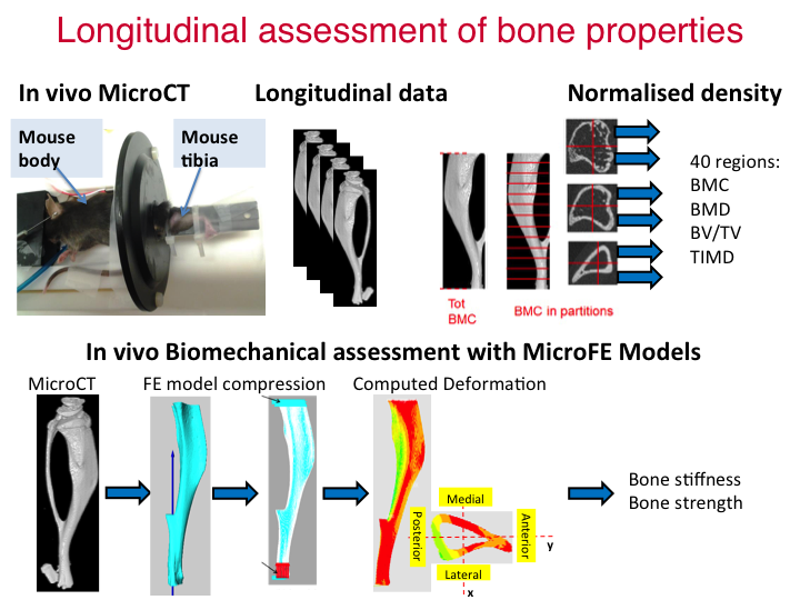
The MouseTibia service provides measurements of densitometric properties in 40 different subregions of the mouse tibia and estimates of the stiffness and strength of the mouse tibia, using a combination of longitudinal micro computed tomography (microCT) and micro finite element (microFE) models. A user uploads a series of microCT datasets from different mice at different time points, and receives back a set of values that characterise the density and mechanical properties for each bone under compression.
The service operates in three steps:
First: the provided images at the different time points and from the different mice are placed in the same reference system by using rigid registration (after having chosen a reference dataset at baseline, if not otherwise indicated the first dataset will be used as reference). The length of each tibia is then calculated and each tibia is cropped below the proximal growth plate for 80% of its length.
Second: an automatic algorithm is run in order to convert the images into equivalent Tissue Mineral Density (TMD). This step assumes that the user has provided the densitometric calibration for each image (if not indicated otherwise, the parameters from the DICOM header will be used to perform the densitometric calibration). Afterwards the script automatically divides the tibia into 10 sections with the same length (8% of the original length of the tibia, called C01 to C10 with C01 the most proximal and C10 the most distal sections) and each section into four quadrants (anterior, medial, posterior and lateral) for a total of 40 compartments. In each compartment the Bone Mineral Content (BMC), Bone Mineral Density (BMD), Bone Volume Fraction (BV/TV) and the Mean Tissue Mineral Density (TMD) are computed.
Third: an automatic algorithm transforms the microCT image into a linear, homogeneous and isotropic microFE model by converting every bone voxel into a linear hexahedral element. Uniaxial compression is applied and the stiffness and strength of the mouse tibia is estimated. These models have been found to be accurate in predicting bone local and structural properties against experimental measurements.
Scientific Background
The mouse tibia is a common site for studying bone remodelling and the effect of bone interventions. In vivo micro-Computed Tomography (microCT) imaging is considered the gold standard for bone imaging and can be applied to measure bone changes over time (Bouxsein et al., 2010). This approach was found to not affect the measurements of bone properties (Oliviero et al., 2019) and dramatically reduce the number of mice needed for studying the effect of diseases (Viceconti and Dall'Ara, 2019). Standard analyses of morphometric parameters are usually performed in two regions of interest (typically 1 mm in length) located at the proximal tibia (trabecular bone) and at the diaphysis (cortical bone). Trabecular morphometric parameters include trabecular bone volume fraction (Tb.BV/TV, [%]), thickness (Tb.Th, [µm]), separation (Tb.Sp, [µm]) and number (Tb.N, [1/mm]). Standard cortical morphometric parameters include total cross-sectional area (Tt.Ar, [mm2]), cortical bone area (Ct.Ar, [mm2]), cortical area fraction (Ct.Ar/Tt.Ar, [mm2/mm2]) and cortical thickness (Ct.Th, [µm]). However, morphometric parameters only provide information about local regions of interest in the tibia.
While these parameters are important to evaluate the effect of interventions on the trabecular and cortical bone, they are not well correlated with the mechanical properties of the mouse tibia (Oliviero et al., 2020). Densitometric properties of the mouse tibia across space and time and mechanical properties of the tibia from longitudinal microCT imaging can provide a complementary assessment of the effect of diseases and treatments in preclinical mouse studies.
Densitometric analysis
Recently a method has been proposed to analyse the whole tibia volume (Lu et al., 2016, Lu et al., 2017), which provides a comprehensive analysis of the whole bone, complementary to the standard morphometric parameters. This method, combined with longitudinal imaging, enables the assessment of the spatio-temporal distribution of the bone densitometric properties.
After rigid registration of the images acquired from different animals or from the same animal at different time points, the attenuation coefficients acquired in the microCT images are converted into tissue mineral density (TMD) using a calibration law based on weekly scans of a densitometric phantom with five insertions. Bone Mineral Content (BMC) in each voxel is calculated as its TMD multiplied by the volume of the voxel. A volume of interest (VOI) is selected below the growth plate, including 80% of the total length and excluding the fibula. Total BMC, TMD and BV/TV are computed in the VOI. The VOI is also divided into ten longitudinal sections (Section1 located at the proximal tibia, Section10 at the distal tibia). For each longitudinal section, the following parameters are measured: bone mineral content (BMC, [mg]), tissue mineral density (TMD, [mgHA/cc]), total bone volume fraction (BV/TV, [%], including both trabecular and cortical bone), total cross-sectional area (TA, [mm2]), bone area (BA, [mm2]), second moment of area in the antero-posterior direction (Ixx, [mm4]) and in the medio-lateral direction (Iyy, [mm4]), polar moment of inertia (Izz, [mm4]). Lastly, each longitudinal section is divided into four quadrants (anterior, posterior, medial and lateral), defined for each cross-section by two perpendicular lines containing its centroid (40 partitions in total), and BMC, TMD and BV/TV are calculated for each partition.
Measurements of the densitometric properties of the mouse tibia obtained with this approach have been proven more repeatable than standard morphometric parameters (Lu et al., 2016) and have been applied to study the effect of bone anabolic interventions such as ovariectomy (Roberts et al., 2019), parathyroid hormone (Lu et al., 2017, Roberts et al., 2020), and passive mechanical loading (alone or in combination with PTH) (Roberts et al., 2020).
microFE analysis
Structural mechanical properties of the tibia and their changes over time are an important endpoint for clinical translation (Viceconti and Dall'Ara, 2019). Micro-Finite Element (microFE) models based on microCT images can be used to estimate the tibia mechanical properties in compression, after proper validation. MicroFE model predictions have been validated against experimental measurements of local displacements obtained with Digital Volume Correlation (R2>0.82 between experimental and predicted local displacements; mean errors for predicted bone strength below 9%) (Oliviero et al., 2018) and experimental measurements of structural properties (mean errors in absolute or normalised strength below 9%) (Oliviero et al., 2020).
The microCT image of the tibia is segmented using a global threshold, calculated as the average of the grey values corresponding to the bone and background peaks in the histogram of the image. A connectivity filter is applied to remove unconnected voxels. A Cartesian mesh is obtained by converting each bone voxel into an 8-noded hexahedral element with isotropic linear elastic material properties (E=14.8 GPa, ν=0.3). Uniaxial compression is simulated by fully constraining the distal surface of the tibia and applying a uniform displacement at the proximal surface. Stiffness ([N/mm]) is calculated as the sum of the reaction forces at the distal end, divided by the applied displacement. Failure load ([N]) is calculated by assuming that the tibia fails when 10% of the nodes reach a critical strain (third principal strain) of -14420 µε (Oliviero et al., 2020).
MouseTibia Service
The MouseTibia Service estimates densitometric and mechanical properties from a microCT image of a mouse tibia.
A job is created by clicking on 'New Job'. This will generate a form in which a microCT image can be selected for the specimen to be analysed. Files can be selected either from Google Drive or your Desktop. A Sheffield University Google account is required to access this service. When you use the Google Drive file picker for the first time this website will ask your permission to add the MouseTibia app to your Google Drive account. You can remove this app at any time in Google Drive by going to:
Settings > Managing Apps > Options > Disconnect from Drive
The user needs to upload a 3D microCT scan as a stack of 2D DICOM images, saved in a compressed .zip folder. Our recommended scan protocol can be found at 'Mouse Tibia Recommended Scan Protocol.docx'. Voxel size and calibration parameters for converting grey levels into TMD are read from the header of the DICOM files. Alternatively, the user can manually input the parameters values in the form, which will overwrite those present in the header. These parameters represent:
- Voxel size [mm]
- Rescale intercept: parameter 0028_1052 in standard Scanco header
- Rescale slope: parameter 0028_1053 in standard Scanco header
- Scale factor: parameter 0029_1000 in standard Scanco header
- Slope of the calibration curve: parameter 0029_1004 in standard Scanco header
- Intercept of the calibration curve: parameter 0029_1005 in standard Scanco header
Once the data have been properly uploaded, an expert operator will process them. The operator will align each image to a reference system. The longitudinal axis of the tibia is aligned to the z axis of the global reference system, the antero-posterior direction is aligned to the x axis and the medio-lateral direction to the y axis. This operation is done with a rigid registration operation, using an aligned tibia as reference. The service also offers the opportunity to upload multiple longitudinal scans of the same bone over time. In this case, the first scan will be used as reference for the alignment of the images acquired at subsequent time points. Lastly, the operator selects a volume of interest (VOI) including the 80% of the total length, starting below the proximal growth plate and excluding the fibula. The user will receive the outputs in a Google Drive folder. For each tibia the following result files are provided:
- Histogram.tif: Histogram (frequency plots) of TMD distribution.
- FE_analysis.xslx: Excel file with estimated structural properties.
- Ten images of binary cross-sections (corresponding to the longitudinal sections, where Section1 corresponds to the proximal tibia, Section10 to the distal tibia).
- For each cross-section, four binary images representing the four sectors (anterior, posterior, medial, lateral).
- Three views of the spatial distribution of first principal strain.
- Histogram_first.tif: Histogram (frequency plots) of first principal strain.
- Three views of the spatial distribution of third principal strain.
- Histogram_third.tif: Histogram (frequency plots) of third principal strain.
- Report in PDF format with a summary of the results (see example in the documents).
The MouseTibia Service
About the service
Detailed information regarding the service and the modelling pipeline can be accessed through these documents. Interested clients would need to provide the following information:
- MicroCT image of the whole mouse tibia for the whole dataset
- Calibration parameters for each image
Please refer to the documents for specifications of the recommended microCT protocol and contact us for clarifications and planning the study.
References
BOUXSEIN, M. L., BOYD, S. K., CHRISTIANSEN, B. A., GULDBERG, R. E., JEPSEN, K. J. & MÜLLER, R. 2010. Guidelines for assessment of bone microstructure in rodents using micro–computed tomography. Journal of Bone and Mineral Research, 25, 1468-1486.
LU, Y., BOUDIFFA, M., DALL'ARA, E., BELLANTUONO, I. & VICECONTI, M. 2016. Development of a protocol to quantify local bone adaptation over space and time: Quantification of reproducibility. Journal of Biomechanics, 2095 - 2099.
LU, Y., BOUDIFFA, M., DALL'ARA, E., LIU, Y., BELLANTUONO, I. & VICECONTI, M. 2017. Longitudinal effects of Parathyroid Hormone treatment on morphological, densitometric and mechanical properties of mouse tibia. Journal of the Mechanical Behavior of Biomedical Materials, 75, 244-251.
OLIVIERO, S., GIORGI, M. & DALL'ARA, E. 2018. Validation of finite element models of the mouse tibia using digital volume correlation. Journal of the Mechanical Behavior of Biomedical Materials, 86, 172-184.
OLIVIERO, S., OWEN, R., REILLY, G. C., BELLANTUONO, I. & DALL'ARA, E. 2020. Optimization of the failure criterion in micro-Finite Element models of the mouse tibia for the non-invasive prediction of its failure load in preclinical applications. Journal of the Mechanical Behavior of Biomedical Materials, 104190.
ROBERTS, B. C., ARREDONDO CARRERA, H. M., ZANJANI-POUR, S., BOUDIFFA, M., WANG, N., GARTLAND, A. & DALL'ARA, E. 2020. PTH(1–34) treatment and/or mechanical loading have different osteogenic effects on the trabecular and cortical bone in the ovariectomized C57BL/6 mouse. Scientific Reports, 10, 8889.
ROBERTS, B. C., GIORGI, M., OLIVIERO, S., WANG, N., BOUDIFFA, M. & DALL'ARA, E. 2019. The longitudinal effects of ovariectomy on the morphometric, densitometric and mechanical properties in the murine tibia: A comparison between two mouse strains. Bone, 127, 260-270.
VICECONTI, M. & DALL'ARA, E. 2019. From bed to bench: How in silico medicine can help ageing research. Mechanisms of Ageing and Development, 177, 103-108.